


What is a retina?
The retina is the thin inner lining of the eyeball. It acts like the film in a camera to “make the picture”. Tiny light-sensitive cells pick up the signals of the world around us and transmit them via the optic nerve to the brain. Because of the way the light falls, the image on the retina is upside-down. Fortunately for us, however, the brain quickly re-invents the picture or else we would all be walking on our hands! Doing all the hard and vitally important work that it does, the retina needs a lot of energy from the body. 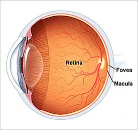 Consequently it has its own little system of blood vessels which have no connections to other vessels. This means that another part of the body cannot steal the retina’s blood but conversely, when a small retinal artery is blocked, severe damage to the retina occurs as there is no blood to steal from anywhere else! When one of the small vessels or their branches becomes blocked sudden loss of vision can occur. No time should be lost in seeing your ophthalmologist because, in the case of a small artery blockage, immediate treatment can sometimes be successful. Your ophthalmologist will probably also want you to undergo some systemic investigations as diseases such as high blood pressure, diabetes and others may be implicated.
Another important fact is that the little light-sensitive cells, which are divided into those that work in the dark (called rods) and those that work in the light (called cones) are constantly breaking down and re-building. When things go wrong with this system retinal degeneration occurs. The cones are mostly concentrated in the central or “bull’s eye” area (called the macula) which we use for clear vision and reading while the rods, which are more concentrated in the periphery, are needed for night vision.
An important problem affecting the macula occurs as the eye (and the patient!) ages. Slow deterioration in this area makes reading and fine close work difficult. Tiny blood vessels may develop and cause distortion of vision or even bleeding. Your ophthalmologist may recommend special photographs or laser treatment to the back of your eye. It is important to remember that the treatment is aimed at preventing further deterioration only. Unfortunately, the opposite eye may also become involved over a period of years. We call this condition Age Related Macular Degeneration. Considerable research is being done and surgical techniques being developed which may occasionally be helpful.
Inherited disorders may cause macular deterioration in much younger patients but one of the commoner degeneration’s is Retinitis Pigmentosa which, especially in the early stages, affects the rods and causes night blindness and “tunnel vision”. Genetic research has helped us to identify some of the faulty genes and the race is on to find a cure.
Another serious condition which may affect the retina is retinal detachment. This means that the retina has come loose from the back of the eye much like wallpaper peeling of a wall! This usually happens when a tear or hole develops in the retina allowing fluid from inside the eye to seep under the retina. The first thing that the patient may notice is flashes of light or a shower of little dots called “floaters”. It is important to see your ophthalmologist if you notice these symptoms, especially if you are short sighted or anyone else in your family has had a retinal detachment. The next thing that happens is that a “curtain” or shadow comes over the vision. When surgery is offered this may be a procedure involving mainly the outside of the eye or may be internal with the injection of a gas bubble or in very severe cases a special oil called silicone oil. The most important thing in the successful treatment of retinal disease is to present as early as possible to your ophthalmologist if you notice any of the symptoms we have mentioned especially if you have a family history of retinal disorders.